Infiltration réalisée sous contrôle radioscopique ou échographique par le médecin radiologue, à but antalgique.
Toutes les articulations peuvent bénéficier d’une infiltration.
L’acte est quasiment indolore (une anesthésie étant couplée à l’injection).
La substance injectée peut être un corticoïde local, parfois une visco-supplémentation (genou).
Après asepsie cutanée stricte, on injecte le principe actif après avoir opacifié l’articulation avec un produit de contraste iodé.
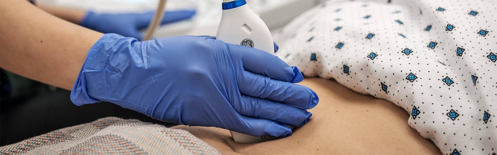
Échographie :
Il est important de rapporter ses anciens examens.
Pour les échographies abdominales il est nécessaire d’être à jeun de 4 heures (à l’exception des patients diabétiques pour lesquels une petite collation peut être envisagée).
Pour les échographies de la vessie et les échographies pelviennes, il est nécessaire d’avoir la vessie pleine et de boire avant l’examen : n’hésitez pas à demander à boire en salle d’attente !
Radiographie :
Vous devez rapporter vos anciens clichés (concernant le même organe).
Comme pour tout examen utilisant les rayons X, vous devez signaler si vous êtes enceinte ou désireuse de l’être (arrêt de la contraception).
Parfois, une vérification de la coagulation sanguine est nécessaire : une prise de sang sera alors effectuée dans un laboratoire d’analyses médicales.
Une ordonnance vous sera adressée ou remise avec votre convocation.
Avant l’examen, vous devez nous prévenir si vous souffrez d’un diabète, d’une anomalie de la coagulation ou d’un antécédent d’allergie médicamenteuse.
Il est nécessaire de consulter un médecin en cas de douleurs, de rougeurs locales ou de fièvre apparaissant dans les jours qui suivent l’infiltration.
Le repos de l'articulation pendant 48H est conseillé .
Technique d’imagerie en coupe utilisant les ultrasons (réflexion des sons sur les tissus et effet Doppler sur les liquides en mouvement).
Il n’y a pas de contre indication, ni d’effets secondaires connus.
Les images sont analysées en temps réel par le radiologue, au chevet du patient. Technique idéale pour l’exploration des parties molles tels les viscères abdominaux, les seins, la thyroïde, les articulations (tendons et ligaments), la prostate.
Couplée à l’effet Doppler elle permet également l’étude des vaisseaux sanguins.
L’échographie permet également de guider des prélèvements diagnostics ou des gestes thérapeutique sous contrôle de l’imagerie tels des infiltrations, biopsies mammaires, biopsies hépatiques...
Il s’agit de la plus ancienne technique d’imagerie médicale, utilisée depuis le début du 20ème siècle.
Technique d’imagerie en projection utilisant les rayons X.
Les doses délivrées sont très faibles, parfois proche de l’irradiation naturelle.
Elles restent utilisées en pratique courante pour l’étude des os, des articulations et des poumons et de manière plus anecdotique des sinus et des dents (panoramique dentaire), ainsi que pour la recherche de calculs urinaires au niveau de l’abdomen (ASP).